IMAgenGINE MRI (Smart Diffusion/Perfusion Post-Processing Tool) Download 2026
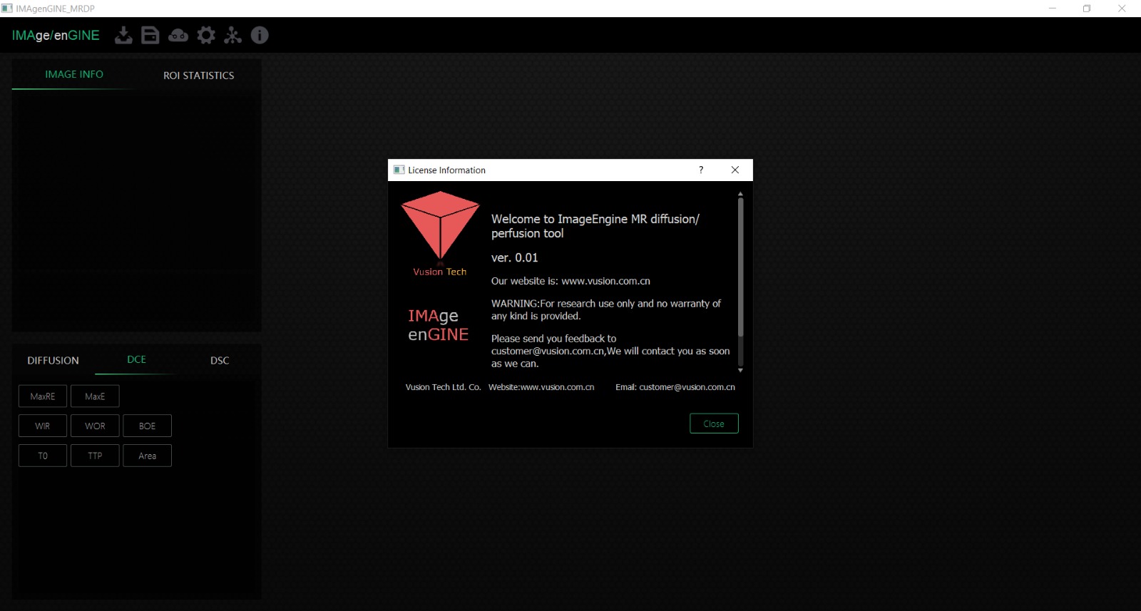
Download the IMAgenGINE MRI (Smart Diffusion/Perfusion Post-Processing Tool) Download 2026 from this link…
![]()
Summary
IMAgenGINE MRI provides customized Medical Imaging Software designed to fit directly into a clinical workflow or your own platform product. It supports advanced functions including MRI diffusion modeling, T1/T2/Permeability quantitative mapping, cardiac function estimation, and other 2D/3D medical imaging post‑processing capabilities such as segmentation, registration, volume rendering, MPR, and more.
These tools help clinicians and researchers analyze complex MRI data effectively and efficiently, enabling detailed assessment of anatomical structures and functional parameters. The Software’s modular design means it can be tailored to the specific needs of different medical departments or research environments.
Core Capabilities: Diffusion and Quantitative MRI Mapping
In advanced medical imaging, diffusion MRI techniques are widely used to examine tissue structure by measuring water movement across tissues, producing parametric maps such as apparent diffusion coefficient (ADC) maps and other derived metrics that help interpret microstructural features of anatomy and pathology. Quantitative mapping methods like T1 maps and T2maps facilitate assessments of tissue characteristics without contrast agents, providing vital data for clinical decision‑making in areas such as tumor evaluation and brain imaging. These quantitative tools, often integrated directly within MRI post‑processing software, serve to enhance the diagnostic capability of standard MRI scanning protocols by offering additional layers of informational depth.
Enhanced Visualization and Post‑Processing Techniques
Modern imaging software supports rich visualization capabilities that go beyond simple image viewing. Tools like 3D rendering and volume visualization enable clinicians to see anatomical structures in three dimensions, providing clearer insights into complex regions of interest. Advanced segmentation and registration features allow the delineation and alignment of areas within medical images, such as isolating tumor boundaries or aligning multi‑modal data from different scan sequences. These post‑processing techniques are essential for creating accurate 3D representations and for supporting measurements, simulation, and planning workflows in both clinical and research settings.
Cardiac MRI Processing and Functional Analysis
Cardiac MRI software modules focus on the heart’s anatomy and function, offering tools to characterize cardiac motion and physiological parameters. For example, cardiac quantitative mapping allows analysis of features such as T1 and T2 relaxation times across myocardial segments to assess tissue health and perfusion. These features are supported through interactive visualization and manual or semi‑automated editing, enabling detailed evaluation of cardiovascular conditions and dynamic heart function. The integration of these advanced MRI cardiac capabilities into post‑processing platforms aids cardiologists and radiologists in tracking disease progression and guiding treatment.
Efficiency Through Integration and Enhanced Workflow Support
Medical imaging platforms often integrate with standardized file formats like DICOM, enabling seamless transfer and handling of images from different sources. Tools such as Medical Imaging Toolbox support visualization, registration, and segmentation across 2D and 3D data while enabling additional capabilities like labeling and preprocessing to improve image quality and analysis workflows.
These integrated systems help reduce manual steps, increase efficiency, and support end‑to‑end workflows from image acquisition through post‑processing and reporting. Such architectures are crucial for ensuring that imaging data is processed accurately and consistently across clinical contexts, from routine diagnostic scans to complex research studies.
Advanced Analysis Support for Clinical Utility
Beyond visualization and mapping, advanced imaging platforms increasingly incorporate analytical tools that enable clinicians to derive meaningful quantitative parameters from MRI data. This includes segmentation of anatomical regions, calculation of parametric maps, and extraction of metrics that assist in disease characterization. The use of segmentation and registration algorithms, ranging from manual adjustments to sophisticated automatic methods, supports both research and patient care by identifying precise anatomical features and changes over time. These analytical layers built into MRI processing systems provide clinicians with additional context and measurements that enhance diagnosis, monitoring, and treatment planning.
Integration of Cutting‑Edge Features
Tools that support diffusion imaging, quantitative mapping, 3D visualization, and advanced segmentation are not just for research; they play a vital role in everyday clinical decision‑making. For example, diffusion parametric maps and quantitative assessments help radiologists detect and classify lesions, while 3D reconstructions assist surgeons and clinicians in planning interventions.
By bringing together these diverse capabilities into a unified platform like IMAgenGINE MRI, medical professionals gain a powerful environment that streamlines workflows from image acquisition to interpretation and reporting. This seamless integration enhances clinical confidence and supports better patient outcomes across specialties.
Supporting Personalized Imaging Needs
The ability to tailor MRI software modules to specific clinical or research requirements adds significant value. Customized solutions can include specialized modules for brain, cardiac, or tumor imaging, each optimized for particular tasks such as diffusion modeling or functional mapping. Customizable workflows also facilitate integration with hospital information systems and research pipelines, ensuring that imaging software works in harmony with existing data infrastructures and clinical requirements. This flexibility helps institutions adopt tools that align with their unique diagnostic and research goals, improving efficiency and the utility of MRI data.
The Future of Medical Imaging Post‑Processing
The landscape of IMAgenGINE MRI software continues to evolve, with emerging technologies pushing the boundaries of what is possible in medical imaging. Advanced segmentation and registration algorithms, enhanced visualization techniques, and quantitative analysis tools are being integrated into next‑generation platforms that aim to provide clinicians with deeper insights and faster workflows.
These developments promise to improve diagnostic accuracy and support personalized patient care through more detailed, efficient, and interpretable imaging data. Platforms like IMAgenGINE MRI represent this future by combining state‑of‑the‑art imaging post‑processing features with flexible deployment options that fit a variety of clinical and research needs.
Our Paid Service
If you want to Purchase Cracked Version / KeyGen Activator /License Key
Contact Us on our Telegram ID :
Join Us For Update Telegram Group :
Join Us For Update WhatsApp group:
Crack Software Policies & Rules:
Lifetime Activation, Unlimited PCs/Users,
You Can test through AnyDesk before Buying,
And When You Are Satisfied, Then Buy It.